Two-Photon Microscopy
We use two-photon microscopy to image fluorescence from genetically encoded sensors of neural activity. This allows us to gather visual responses from retinal neurons ex vivo or centrally located visual neurons in vivo

Mesoscale Calcium Imaging
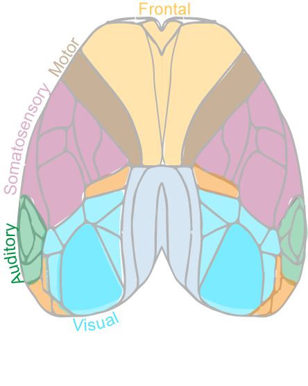
Widefield epifluorescence imaging lets us image neural activity sensors and neurotransmitter sensors across the entire dorsal cortex to see how cortical areas activate and signal to one another while mice perform visually-guided behaviors
Mouse Genetics
The lab has a range of mouse lines that allow us to selectively mark, monitor, and manipulate neurons throughout the early stages of the visual pathway (ie: retina, LGN, visual cortex)

Fiber Photometry
We use fiber photometry to collect neural signals from deep-brain structures and nuclei while mice perform visually guided behaviors

Optogenetics
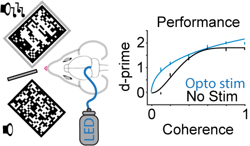
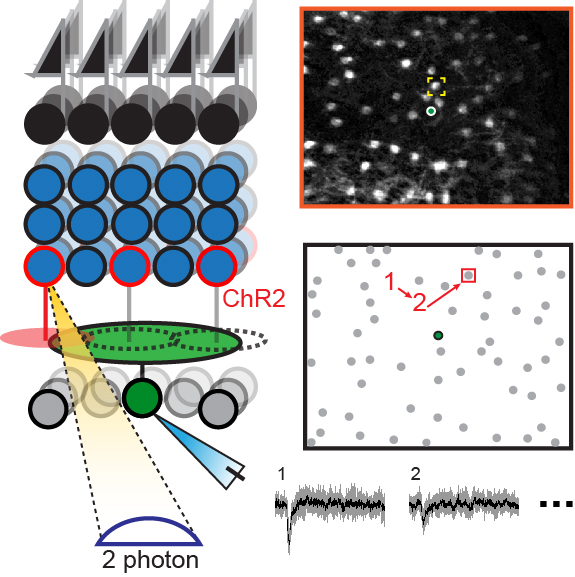
Optogenetic tools allow us to control neural activity with light and permit mapping of functionally connected neurons ex vivo and tests for causal links between candidate brain regions and attentional/perceptual behaviors
Electrophysiology
We are using optogenetics together with patch clamp electrophysiology to map functional connectivity between neurons in retinal explants or dLGN and cortical slices. By relating these patterns to cell-type and by studying how these patterns arise during development we are trying to understand how neurons connect specifically to one another.
Innate and learned Behavior
We use several assays that probe both innate and learned visually-guided behaviors. A learned behavioral task in which mice detect the a 3-bar grating emerging from checkerboard noise is used in several of our studies on visual attention and perception
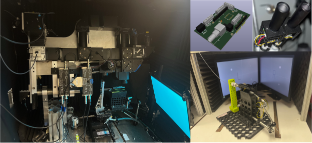
NeuroTechnology
Developing custom microscopes, electronics, behavioral apparatus, and other instrumentation is central to our approach. Our machine shop is equipped with standard tools (band saw, drill press), 3D printers, micro mill, and a laser cutter.